تم الحل ✓
categoryالفيزياء
schoolبكالوريوس
event_available2026-07-14
السؤال
Transcribed Image Text:
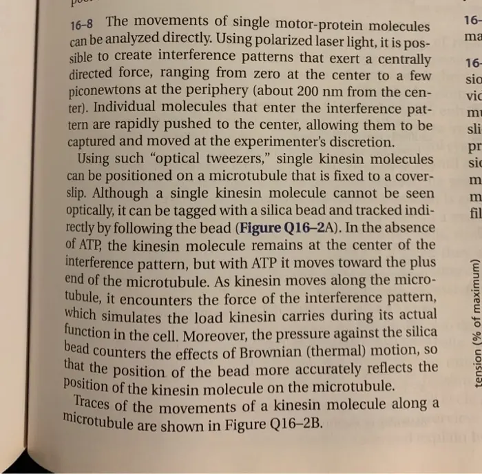
16-8 The movements of single motor-protein molecules
can be analyzed directly. Using polarized laser light, it is pos-
sible to create interference patterns that exert a centrally
directed force, ranging from zero at the center to a few
piconewtons at the periphery (about 200 nm from the cen-
ter). Individual molecules that enter the interference pat-
tern are rapidly pushed to the center, allowing them to be
captured and moved at the experimenter's discretion.
Using such "optical tweezers," single kinesin molecules
can be positioned on a microtubule that is fixed to a cover-
slip. Although a single kinesin molecule cannot be seen
optically, it can be tagged with a silica bead and tracked indi-
rectly by following the bead (Figure Q16-2A). In the absence
of ATP, the kinesin molecule remains at the center of the
interference pattern, but with ATP it moves toward the plus
end of the microtubule. As kinesin moves along the micro-
tubule, it encounters the force of the interference pattern,
which simulates the load kinesin carries during its actual
function in the cell. Moreover, the pressure against the silica
bead counters the effects of Brownian (thermal) motion, so
that the position of the bead more accurately reflects the
position of the kinesin molecule on the microtubule.
Traces of the movements of a kinesin molecule along a
microtubule are shown in Figure Q16-2B.
16-
ma
16-
sia
vic
mu
sli
pr
sic
m
m
fil
tension (% of maximum)
(A) EXPERIMENTAL SETUP
silica
bead
microtubule
distance (nm)
(B) POSITION OF KINESIN
138
80-
trace 1
60-
40-
to s
kinesin
20.
0-
0
2
6
time (seconds)
8
Figure Q16-2 Movement of kinesin along a microtubule (Problem
16-8). (A) Experimental setup with kinesin linked to a silica bead,
moving along a microtubule. (B) Position of kinesin (as visualized by
position of silica bead) relative to center of interference pattern, as a
function of time of movement along the microtubule. The jagged
nature of the trace results from Brownian motion of the bead.
A. As shown in Figure Q16-2B, all movement of kinesin is
in one direction (toward the plus end of the microtubule).
What supplies the free energy needed to ensure a unidirec-
tional movement along the microtubule?
B. What is the average rate of movement of kinesin along
the microtubule?
C. What is the length of each step that a kinesin takes as it
moves along a microtubule?
D. From other studies it is known that kinesin has two
globular domains that each can bind to ẞ-tubulin, and that
kinesin moves along a single protofilament in a micro-
tubule. In each protofilament the ẞ-tubulin subunit repeats
at 8-nm intervals. Given the step length and the interval
between ẞ-tubulin subunits, how do you suppose a kinesin
molecule moves along a microtubule?
E. Is there anything in the data in Figure Q16-2B that tells
you how many ATP molecules are hydrolyzed per step?
check_circle الجواب — حل مفصل خطوة بخطوة
hourglass_top
🔒
الحل الكامل متاح للمشتركين
اشترك في أرشيف الأسئلة لعرض هذا الحل وآلاف الحلول المفصلة خطوة بخطوة من معلمين معتمدين.