تم الحل ✓
categoryأحياء
schoolبكالوريوس
event_available2026-07-15
السؤال
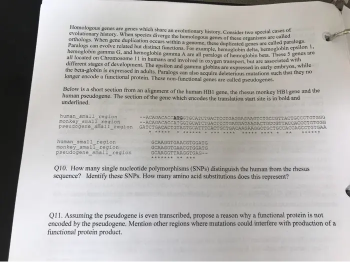
Transcribed Image Text:
Homologous genes are genes which share an evolutionary history. Consider two special cases of
evolutionary history. When species diverge the homologous genes of these organisms are called
orthologs. When gene duplication occurs within a genome, these duplicated genes are called paralog
Paralogs can evolve related but distinct functions. For example, hemoglobin delta, hemoglobin epsilon 1.
hemoglobin gamma G, and hemoglobin gamma A are all paralogs of hemoglobin beta. These 5 genes are
all located on Chromosome 11 in humans and involved in oxygen transport, but are associated with
different stages of development. The epsilon and gamma globins are expressed in early embryos, while
the beta-globin is expressed in adults. Paralogs can also acquire deleterious mutations such that they no
longer encode a functional protein. These non-functional genes are called pseudogenes.
Below is a short section from an alignment of the human HB1 gene, the rhesus monkey HB1 gene and the
human pseudogene. The section of the gene which encodes the translation start site is in bold and
underlined.
human_small_region
monkey small_region
pseudogene_small_region
human_small_region.
monkey small region
pseudogene_small_region
--ACAGACACCATGGTGCATCTGACTCCTGAGGAGAAG?
CCTGTGGG
--ACAGACACCATGGTGCATCTGACTCCTGAGGAGAAGACTGCCGTTACCACCCTGTGGG
GATCTGACACTGTAGTGCATTTCACTGCTGACAAGAAGGCTGCTGCCACCAGCCTGTGAA
*******
GCAAGGTGAACGTGGATG
GCAAGGTGAACGTGGATG
GCAAGGTTAAGGTGAG--
*****
Q10. How many single nucleotide polymorphisms (SNPs) distinguish the human from the rhesus
sequence? Identify these SNPs. How many amino acid substitutions does this represent?
Q11. Assuming the pseudogene is even transcribed, propose a reason why a functional protein is not
encoded by the pseudogene. Mention other regions where mutations could interfere with production of a
functional protein product.
Intron-exon junctions have characteristic nucleotide sequences that are found at the 5' and 3' junctions.
represent the relative occurrence of each nucleotide at each position in the sequence, the larger the letter
the more often it is expected to be observed at that position.
Below are two sequence logos made from the intron-exon junctions of many human genes
(http://weblogo.berkeley.edu/examples.html). The breakpoint junction in both diagrams is between
-1 and 0.
exon | intron
intron exon
0-
2345678
44432123
Q4. Make a list of the first six nucleotides of each intron. Do you see any patterns? Do these patterns
match your expectations?
Q5. Make a list of the last six nucleotides of each intron. Do you see any patterns? Do these patterns
match your expectations?
The branch point sequence of the intron which contains that critical "A" nucleotide generally have the
following sequence "YNCURAY" where Y indicates any pyrimidine, R indicates and purine, and N
indicates any base.
Q6. Can you identify this sequence for the first intron? Mark and label it and write the precise sequence
here. Within the gene, are the two exons flanking this first intron in the same reading frame? Does that
matter? Explain.
Codons consist of 3 nucleotide combinations, but the length of the mRNA (626 nt) shown below divided
by 3 is 208.67 and is greater than the number of amino acids, 147, in the polypeptide.
Q7. Explain the difference and label these differences in your sequence above.
Q8. What are the sequence of nucleotides in the start and stop codons?
Regions between start and stop codons in the same reading frame are called "open reading frames" or
ORFs. Working in groups, identify the start and stop codons in each reading frame. The mRNA sequence
is printed below (hint: you can search for sequences in WORD to make this easy).
Homo sapiens beta globin mRNA
ACAUUUGCUUCUGACACAACUGUGUUCACUAGCAACCUCAAACAGACACCAUGGUGCAUCUGACUCCUGAGGAGAAG
UCUGCCGUUACUGCCCUGUGGGGCAAGGUGAACGUGGAUGAAGUUGGUGGUGAGGCCCUGGGCAGGCUGCUGGUGGU
CUACCCUUGGACCCAGAGGUUCUUUGAGUCCUUUGGGGAUCUGUCCACUCCUGAUGCUGUUAUGGGCAACCCUAAGG
UGAAGGCUCAUGGCAAGAAAGUGCUCGGUGCCUUUAGUGAUGGCCUGGCUCACCUGGACAACCUCAAGGGCACCUUU
GCCACACUGAGUGAGCUGCACUGUGACAAGCUGCACGUGGAUCCUGAGAACUUCAGGCUCCUGGGCAACGUGCUGGU
CUGUGUGCUGGCCCAUCACUUUGGCAAAGAAUUCACCCCACCAGUGCAGGCUGCCUAUCAGAAAGUGGUGGCUGGUG
UGGCUAAUGCCCUGGCCCACAAGUAUCACUAAGCUCGCUUUCUUGCUGUCCAAUUUCUAUUAAAGGUUCCUUUGUUC
CCUAAGUCCAACUACUAAACUGGGGGAUAUUAUGAAGGGCCUUGAGCAUCUGGAUUCUGCCUAAUAAAAAACAUUUA
UUUUCAUUGCAAAAAAAAAAA
AAAAAAAAAA
Q9. Which ORF do you think is most likely to represent the actual coding sequence of this gene? Explain.
Homologous genes are genes which share an evolutionary history. Consider two special cases of
evolutionary history. When species diverge the homologous genes of these organisms are called
orthologs. When gene duplication occurs within a genome, these duplicated genes are called paralogs.
Paralogs can evolve related but distinct functions. For example, hemoglobin delta, hemoglobin epsilon 1.
hemoglobin gamma G, and hemoglobin gamma A are all paralogs of hemoglobin beta. These 5 genes are
all located on Chromosome 11 in humans and involved in oxygen transport, but are associated with
different stages of development. The epsilon and gamma globins are expressed in early embryos, while
the beta-globin is expressed in adults. Paralogs can also acquire deleterious mutations such that they no
longer encode a functional protein. These non-functional genes are called pseudogenes.
Below is a short section from an alignment of the human HB1 gene, the rhesus monkey HB1 gene and the
human pseudogene. The section of the gene which encodes the translation start site is in bold and
underlined.
human_small_region
monkey small region
pseudogene_small_region
human_small_region
monkey small_region
pseudogene_small_region
--ACAGACACCATGGTGCATCTGACTCCTGAGGAGAAGTCTGCCGTTACTGCCCTGTGGG
GATCTGACACTGTAGTGCATTTCACTGCTGACAAGAAGGCTGCTGCCACCAGCCTGTGAA
--ACAGACACCATGGTGCATCTGACTCCTGAGGAGAAGACTGCCGTTACCACCCTGTGGG
*****
GCAAGGTGAACGTGGATG
GCAAGGTGAACGTGGATG
GCAAGGTTAAGGTGAG--
*****
Q10. How many single nucleotide polymorphisms (SNPs) distinguish the human from the rhesus
sequence? Identify these SNPs. How many amino acid substitutions does this represent?
Q11. Assuming the pseudogene is even transcribed, propose a reason why a functional protein is not
encoded by the pseudogene. Mention other regions where mutations could interfere with production of a
functional protein product.
Sickle cell anemia is associated with a single nucleotide difference which results in a non-synonymous
mutation (a change in the nucleotide sequence results in a change to the amino acid encoded). The SNP
changes the encoded amino acid from a glutamic acid (glu - normal) to valine (val-sickle cell). From
the start codon for the large ORF below, can you find the codon for the 7 amino acid (counting the
methionine of the start as the first)?
Q12. What possible codons could the normal glutamic acid be?
Q13. Which is actually found in this sequence? Draw a box around this region.
Q14. What type of mutation is this single nucleotide difference? A transition (Pyrimidine to Pyrimidine or
Purine to Purine), a transversion (Pur to Pyr or Pyr to Pur), an insertion, a deletion...?
Q15. Where is the poly-adenylation ("poly-A") signal, "AAUAAA" located? Draw a box around this
region.
check_circle الجواب — حل مفصل خطوة بخطوة
hourglass_top
🔒
الحل الكامل متاح للمشتركين
اشترك في أرشيف الأسئلة لعرض هذا الحل وآلاف الحلول المفصلة خطوة بخطوة من معلمين معتمدين.